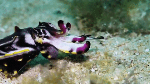
The chromatophores are a sac containing hundreds of thousands of pigment granules and a large membrane that is folded when retracted. There are hundreds of muscles radiating from the chromatophore. These are under neural control and when they expand, they reveal the hue of the pigment contained in the sac. Cuttlefish have three types of chromatophore: yellow/orange (the uppermost layer), red, and brown/black (the deepest layer). The cuttlefish can control the contraction and relaxation of the muscles around individual chromatophores, thereby opening or closing the elastic sacs and allowing different levels of pigment to be exposed. Furthermore, the chromatophores contain luminescent protein nanostructures; there are tethered pigment granules which modify light through absorbance, reflection, and fluorescence between 650 and 720 nm.
In cuttlefish, activation of a chromatophore can expand its surface area by 500%. There may be up to 200 chromatophores per mm2 of skin. In Loligo plei, an expanded chromatophore may be up to 1.5 mm in diameter, but when retracted, it can measure as little as 0.1mm.
Retracting the chromatophores reveals the iridophores and leucophores beneath them, thereby allowing cuttlefish to use another modality of visual signalling brought about by structural coloration.
Iridophores are structures that produce iridescent colors with a metallic sheen. They reflect light using plates of crystalline chemochromes made from guanine. When illuminated, they reflect iridescent colors because of the diffraction of light within the stacked plates. Orientation of the schemochrome determines the nature of the color observed. By using biochromes as colored filters, iridophores create an optical effect known as Tyndall or Rayleigh scattering, producing bright blue or blue-green colors.
https://www.nature.com/scitable/topi...-the-144048968


 Reply With Quote
Bookmarks